
Customised 3D anatomical models
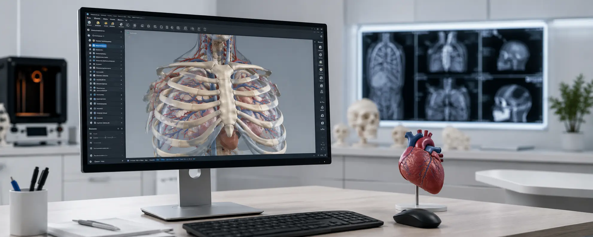
A customised 3D anatomical model is a faithful reproduction of a patient’s anatomical area of interest (organs, bone and tendon structures, muscles, vascular networks, etc.). Unlike standard models, it is designed from data obtained through medical imaging (CT scan, MRI, CBCT), which gives it outstanding accuracy and a high degree of realism..
These models have become a valuable resource in the medical sector. They are used to prepare and rehearse surgical procedures, to train healthcare professionals and for medical research. They are also used to communicate with patients, offering a clear and realistic representation of their anatomy and/or pathology.
Their creation relies on two main stages: the digital segmentation / modelling phase, which turns medical images into usable 3D files, followed by 3D printing, which brings this data to life as a precise physical object, at full scale or adapted to the needs of the project.
How does the
3D manufacturing process work?
Acquisition and modelling
of medical data
The creation of a customised 3D anatomical model begins with the acquisition of data from medical imaging (CT scan, MRI, CBCT, etc.). This information is then processed using specialist software such as Simpleware, which converts the images into a precise, usable digital model (digital twin).
Design
and preparation of the model
Once the model has been generated, it can be refined using digital design tools. This stage makes it possible to adjust the level of detail, add supports or functions, change the scale or target a particular system (bone, organ, muscle). This is where the customisation takes shape, according to clinical or educational needs.
Manufacturing
by 3D printing
The final stage consists of turning the digital model into a physical object through 3D printing. Depending on the application, different materials can be used: plastic, resin, or biocompatible materials for more advanced use. This process makes it possible to quickly produce a realistic, accurate model that healthcare professionals can use straight away.
What are the benefits of 3D models for medical education?
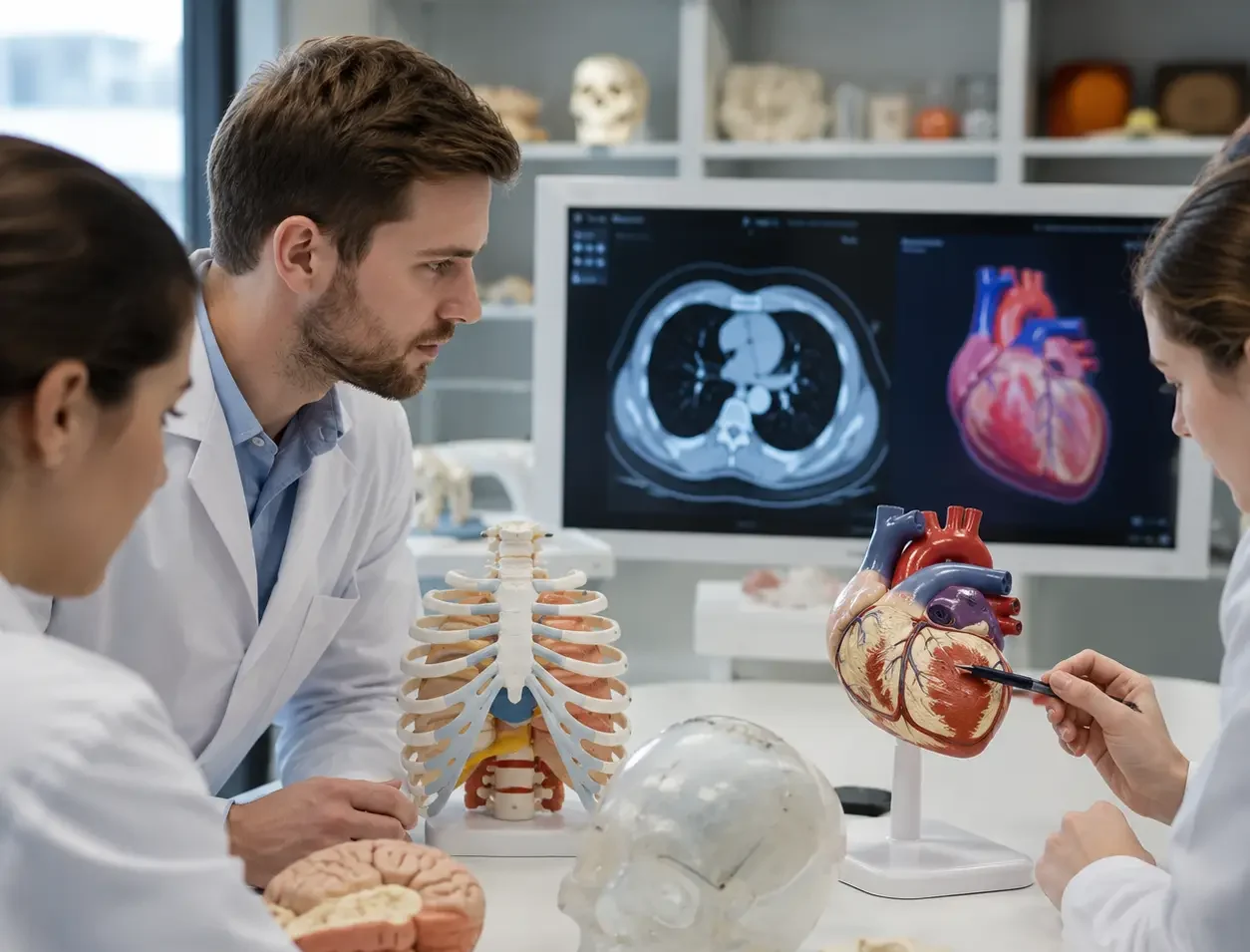
In medical education, 3D anatomical models have become concrete, interactive learning aids:
- They make it possible to simulate procedures and to clearly visualise certain complex pathologies.
- They facilitate communication with patients by making an operation or the workings of an organ easier to understand.
- They complement digital platforms and theory lectures effectively, reinforcing learning through practice.
What types of anatomical models are there?
3D anatomical models come in several forms depending on the medical or educational use:
- Muscular system models
- Skeleton and bone models
- Organ models (heart, liver, lungs, etc.)
- Detailed models for surgery or interventional radiology
- Interactive and customisable models
Which software is used to create 3D anatomical models?
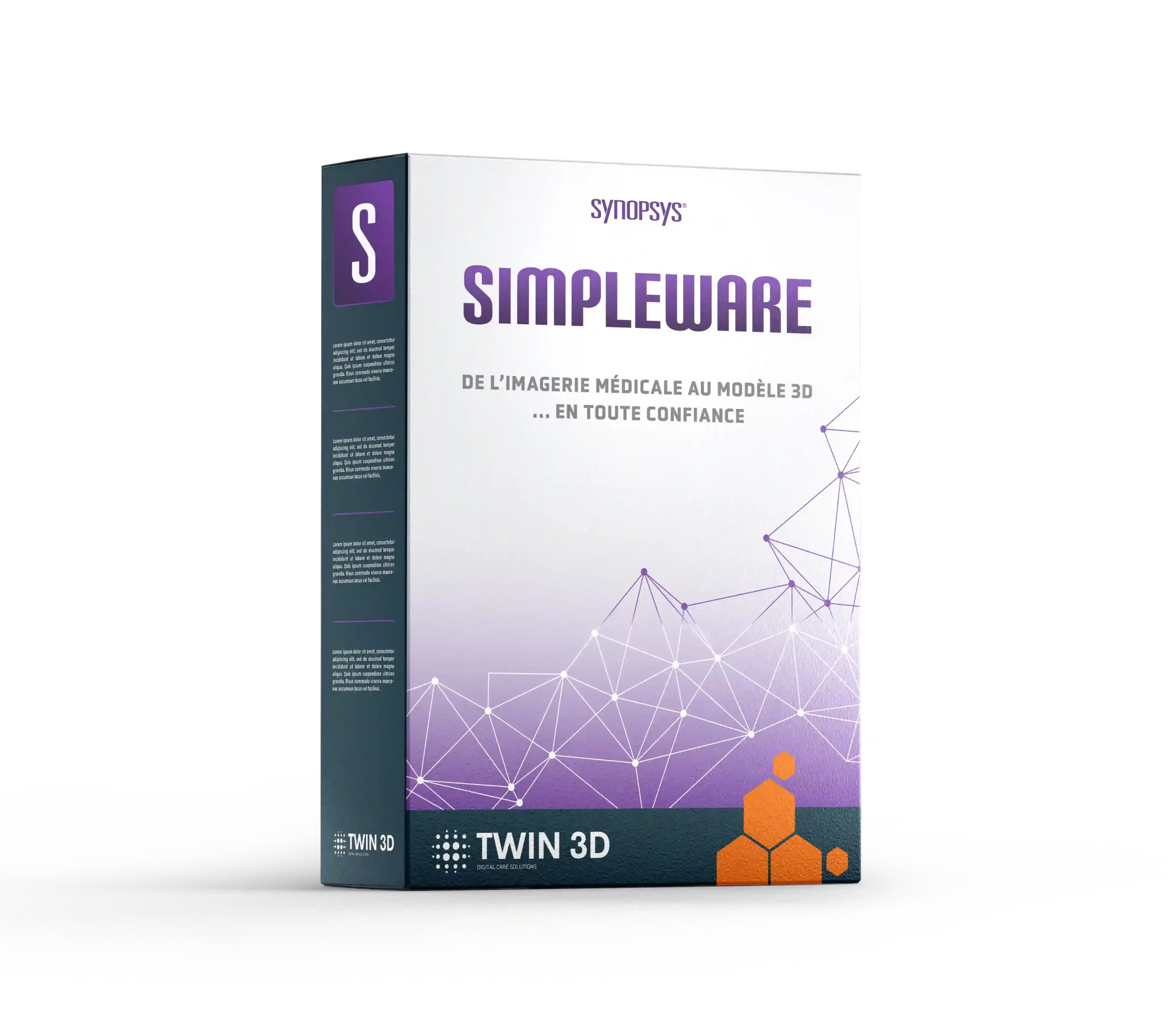
Creating a customised 3D anatomical model requires suitable software. Simpleware is the benchmark for turning medical imaging into a precise digital model.
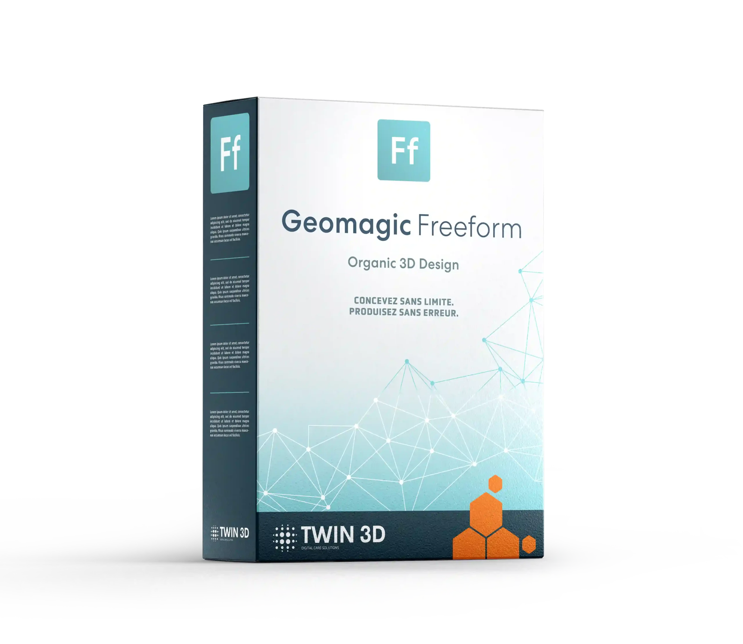
Depending on requirements, CAD tools such as Geomagic Freeform, which are compatible with medical 3D printing, complete the process.
The stages of using an anatomical model with Twin 3D
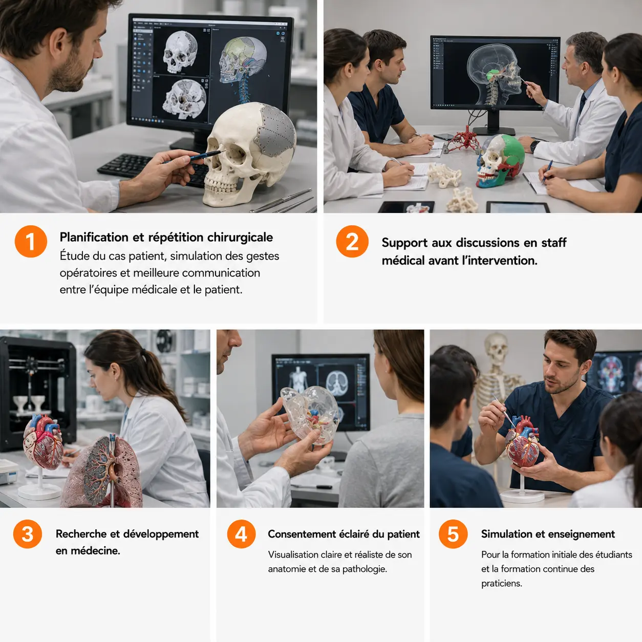
A customised 3D anatomical model can be used at different stages of the patient pathway:
-
1
Surgical planning and rehearsal: patient case study, simulation of surgical procedures and better communication between the medical team and the patient.
-
2
Support for medical staff discussions before the procedure.
-
3
Research and development in medicine.
-
4
Informed patient consent, with a clear and realistic visualisation of their anatomy and pathology.
-
5
Simulation and teaching for the initial training of students and the continuing professional development of practitioners.
Solutions offered by Twin 3D

Specialist software

Robotic arms


Training courses
FAQ
They involve checks on accuracy, strength and, in some cases, sterilisation. Materials must comply with ISO standards (e.g. ISO 10993) and, depending on the case, regulatory requirements (MDR, FDA).
Medical imaging files (CT scan, MRI, CBCT) are anonymised and transferred securely. Compliance with the GDPR and medical confidentiality is mandatory and guaranteed by our software.
It all depends on the intended use: low-cost plastic for teaching, high-precision resin for surgical simulation, biocompatible and sterilisable materials for the operating theatre, or radiopaque materials for use in radiology.
Other medical uses
They trust us









Book a demonstration
Twin 3D supports healthcare professionals in their medical 3D projects. Speak with an expert and discover our solutions during a demonstration.