
3D technologies applied to cardiology
The use of 3D in cardiology opens up new prospects for the management of complex heart conditions. Thanks to digital modelling and additive manufacturing, it is now possible to design anatomical models of the heart and blood vessels, prepare for delicate cardiac surgery and develop innovative medical devices.
At Twin 3D, we work with medical teams and hospital centres to turn cardiac imaging into practical tools: 3D-printed models, surgical guides and digital solutions that support surgical planning and improve patient information and safety.
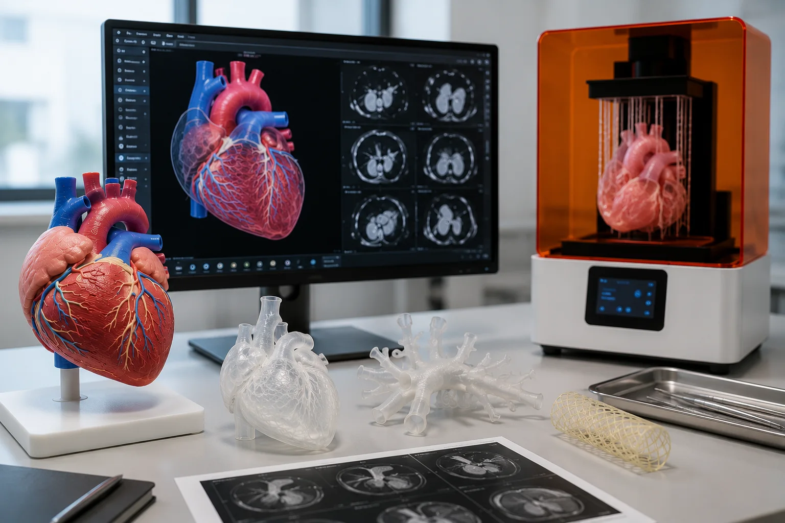
How do 3D technologies help in cardiology?
3D software provides tools for understanding and preparation for cardiologists and cardiac surgeons:
- Better understanding of complex conditions: congenital anomalies, malformations, aneurysms. This new way of visualising anatomical structures and pathologies facilitates multidisciplinary discussions between healthcare professionals (team meetings).
- Surgical simulation: faithful reproduction of the chambers, ventricles and vessels to anticipate the procedure or to simulate fluid flow and behaviour.
- Decision support: 3D modelling of an organ before an operation to choose the most suitable technique.
Communication support: clearer explanation of the treatment to patients and their families to facilitate informed consent.
These printed models enable teams to reduce complications and intraoperative time, and to improve the success rate of cardiac procedures.
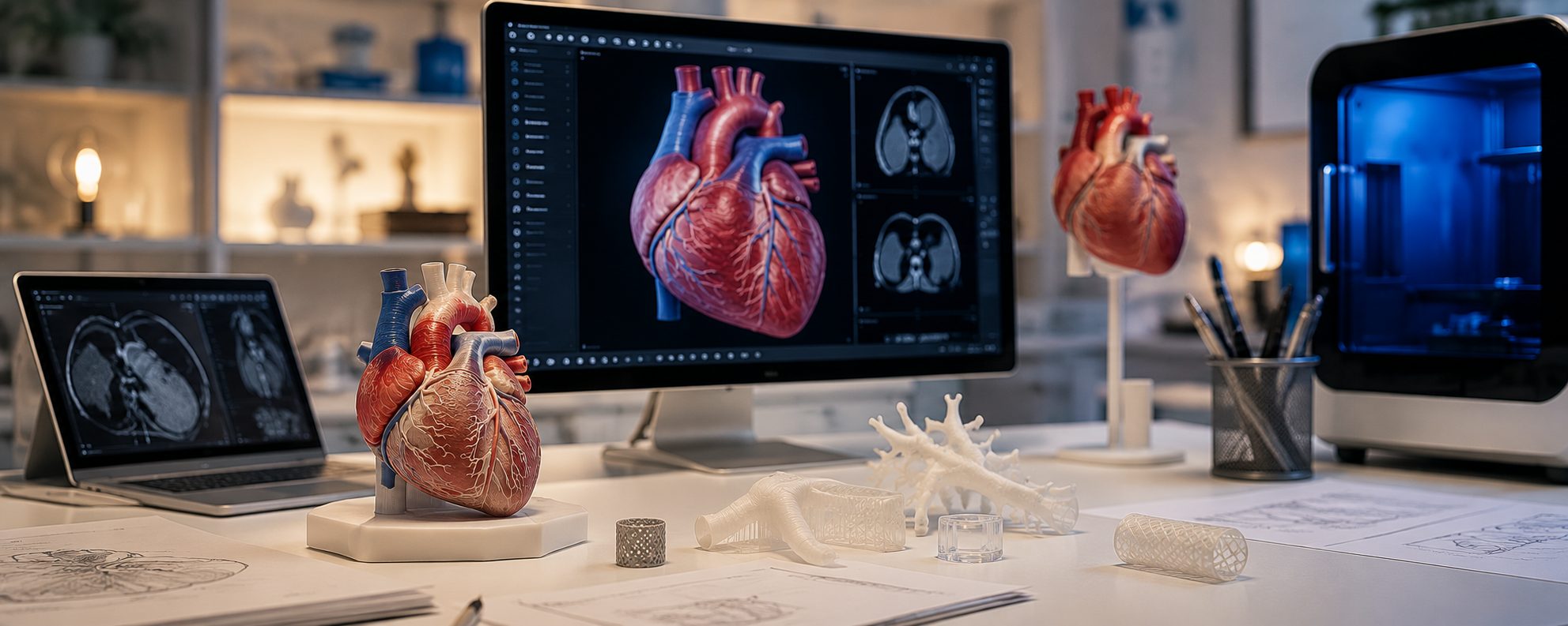
What are the benefits of 3D printing in healthcare?
In cardiology, as in other disciplines, the benefits of 3D printing are many:

High precision
Anatomical models and medical devices.

Speed and efficiency
Shorter planning, reduced operating time.

Training and learning
Realistic teaching tools to train young surgeons.

Personalised medicine
Solutions tailored to the patient's anatomy.

Continuous innovation
Opening the way to bioprinting and the creation of living tissue.
What innovations exist in 3D bioprinting?
Though currently explored mainly in medical research, 3D bioprinting combines bio-inks, cells and tissue engineering. It opens up promising avenues, in particular:
- The design of functional heart valves in the laboratory,
- The printing of cardiac tissue for experimentation,
- The reproduction of complex vascular structures to better understand blood circulation and, in the longer term, the ambition of creating a bioprinted heart for transplantation.
This work remains experimental, but it illustrates how 3D printing could transform cardiology in the years to come.
How do you 3D print a heart?
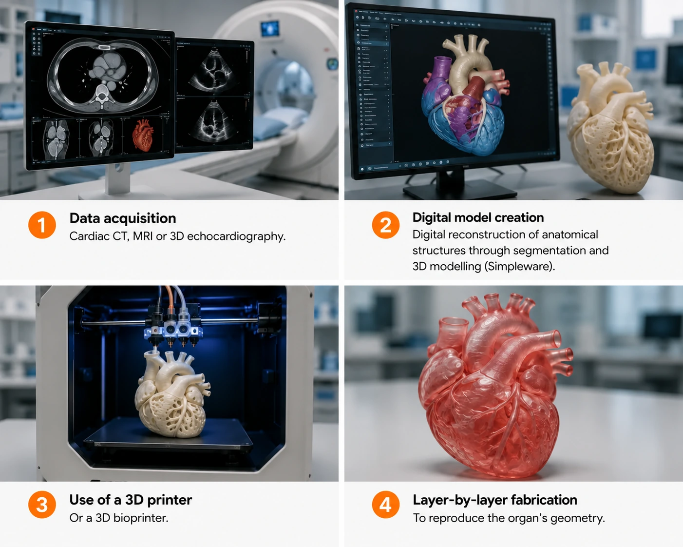
3D printing applied to the heart takes place in several phases:
-
1
Data acquisition: cardiac CT scan, MRI or 3D echocardiography.
-
2
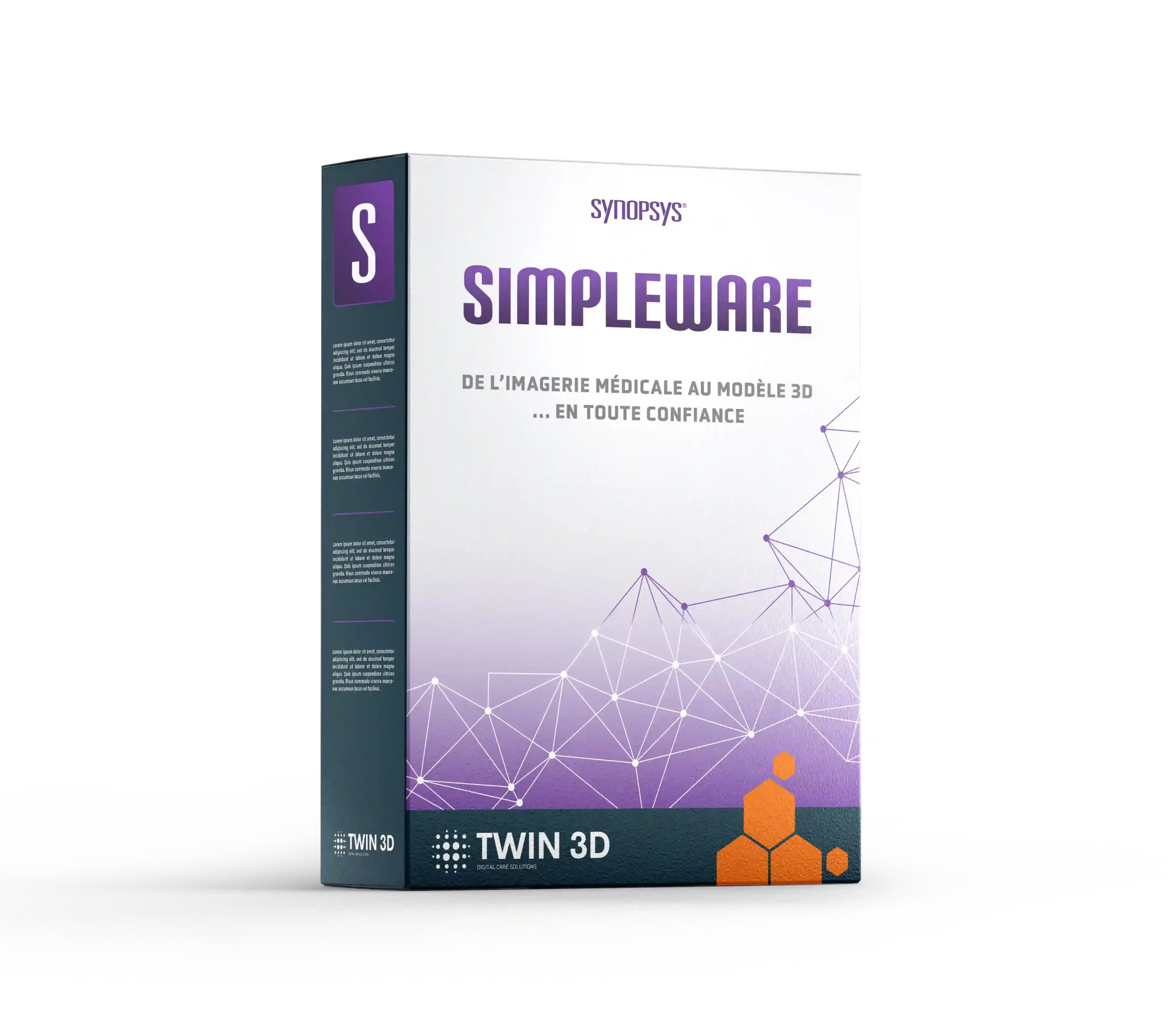
Creation of the digital model: digital reconstruction of anatomical structures through segmentation and 3D modelling (Simpleware)
-
3
Use of a 3D printer or a 3D bioprinter.
-
4
Layer-by-layer fabrication to reproduce the geometry of the organ.
Today, anatomical models of the heart can already be produced for surgical planning, while the creation of a living organ remains at the experimental stage.
What 3D applications are available for cardiology?
3D models in cardiology are varied and meet specific needs:
Printed anatomical models
For simulating procedures or for initial and continuing medical training

3D digital files
Available for research, training (for example via Google Scholar and university databases), multidisciplinary discussions between healthcare professionals or patient information
Complex cardiac structures
Ventricles, atria, coronary vessels.

Paediatric models
To better treat babies and children with malformations.
Solutions offered by Twin 3D
- Certified software for segmentation, modelling, cardiac planning and the design of patient-specific devices.
- Printed anatomical models for surgical preparation and teaching.
- Comprehensive support: installation, training, technical support and clinical follow-up.
Our goal: to give medical teams reliable, advanced tools that they can use fully autonomously to improve the care of cardiology patients.
Solutions offered by Twin 3D
Specialist software

Robotic arms


Training courses
FAQ
Yes. Our software supports standard formats (DICOM, STL, OBJ), ensuring compatibility with cardiac imaging systems and hospital equipment (PACS)
They trust us








Book a demonstration
Twin 3D supports healthcare professionals in their medical 3D projects. Speak with an expert and discover our solutions during a demonstration.