
3D technology and 3D printing applied to radiology
Radiology generates large volumes of clinical images every day (CT, MRI, PET, etc.), often limited to 2D viewing that is difficult to exploit fully. Medical 3D technologies make it possible to turn this data into accurate anatomical models, useful for diagnosis and surgical planning.
Twin 3D supports imaging departments and radiologists with solutions for segmentation, 3D modelling (meshing), additive manufacturing and rapid prototyping, in the service of more personalised and effective medicine.
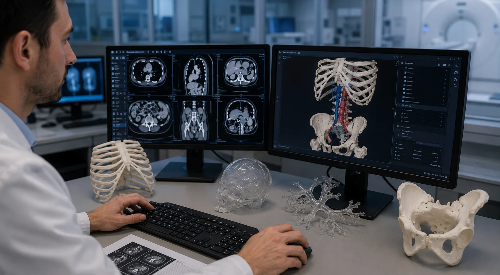
How does 3D printing work in radiology?
The digital workflow relies on a structured process that combines medical imaging and additive manufacturing:
- Data acquisition: the CT scanner, MRI or magnetic resonance imaging system provides high-resolution images in DICOM format.
- Segmentation and 3D modelling: the data is reconstructed into 3D models using modelling software and a multiplanar reconstruction console. This produces accurate anatomical models (organ, tissue, vessels, tumour) tailored to clinical needs.
- Design (CAD): of patient-specific devices or suitable supports for the anatomical models
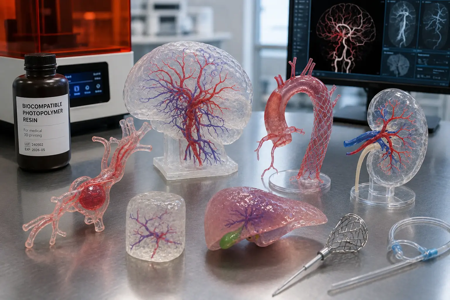
- Additive manufacturing: layer-by-layer printing with biocompatible materials such as resin, polymers, titanium or bioresorbable materials.
- Clinical use: surgical planning, educational simulation, design of customised medical devices or patient information for informed consent and better adherence to treatment.
Result: a better understanding of anatomy, tangible models to anticipate surgical procedures and more personalised medicine.
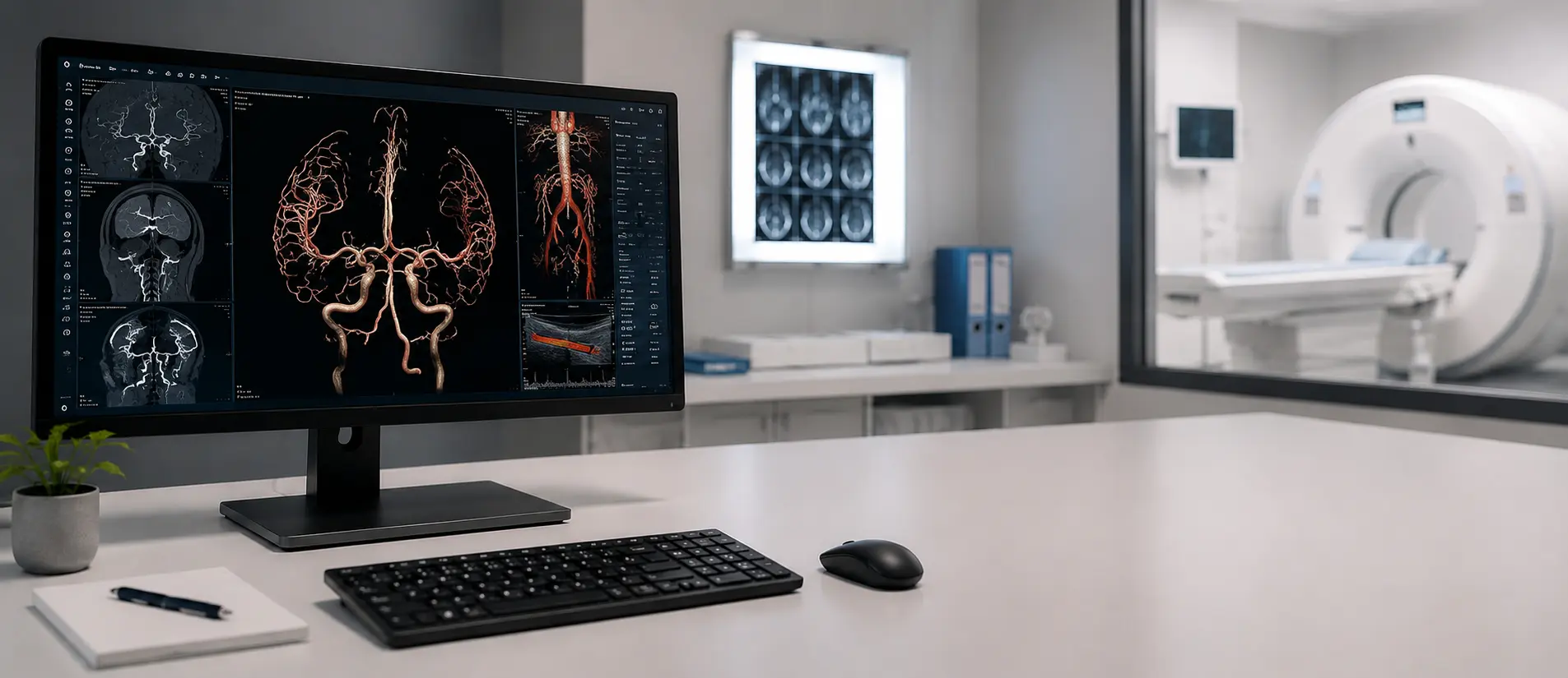
The benefits for radiologists
Integrating 3D printing into radiology brings several concrete benefits:

Easier diagnosis
3D visualisation and exact reproduction of complex structures (kidney, vessels, tumours, etc.) improve interpretation and accuracy.

Personalised medicine
each medical device is custom-made to meet the patient’s specific needs.

Easier communication
reconstructed or printed models serve as an educational aid to explain the pathology to patients and improve their adherence to treatment. They also facilitate multidisciplinary discussions between healthcare professionals (staff meetings).

Time saved for physicians
surgical planning and rapid prototyping help reduce complications and optimise operating time.

Training and research
tangible aids for medical students, hospital teams and clinical research projects.

Practical applications in radiology and healthcare
3D printing in radiology has many applications in the healthcare sector:

Surgical planning
Transfer of accurate models to maxillofacial, oncological or urological surgery departments.

Operative preparation
Anticipation of complex procedures thanks to full-scale anatomical replicas.

Patient-specific devices
Design and manufacture of implants and surgical guides, boluses and immobilisation supports for radiotherapy, or even anatomical models

Research and innovation
Collaboration with hospital teams, such as at Bordeaux University Hospital, to develop new medical practices.
Teaching and simulation
Use of printed models to train residents, radiologists and surgeons.
Which medical devices can be printed?
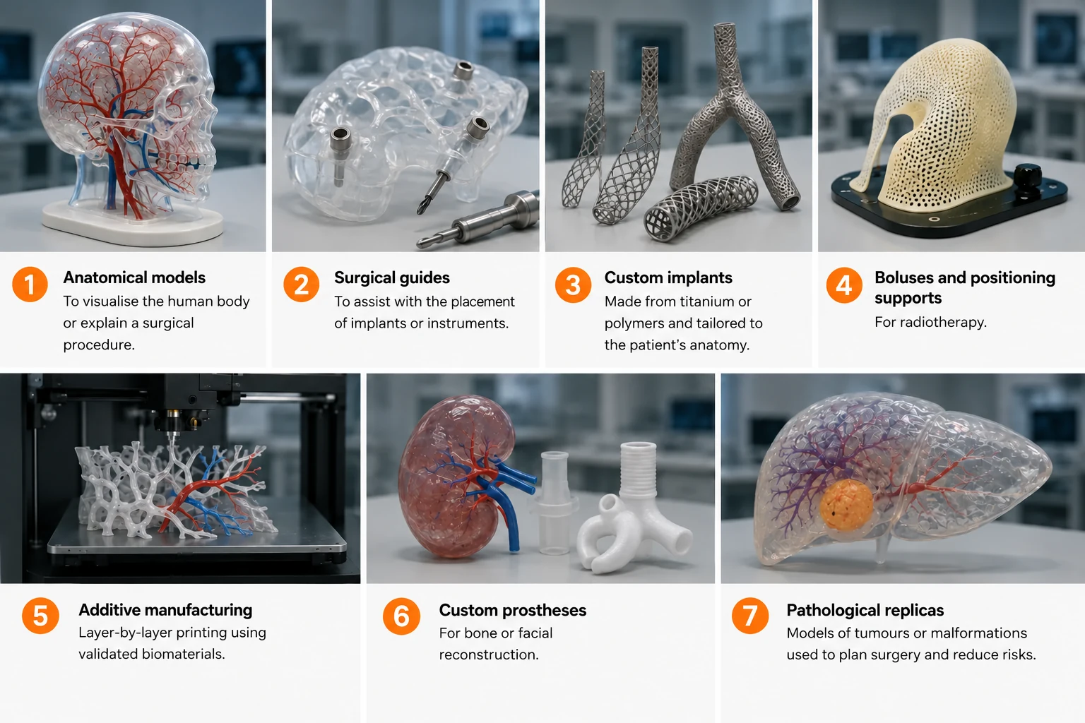
Additive manufacturing makes it possible to produce different types of devices:
-
1
Anatomical models to visualise the human body or explain a procedure.
-
2
Surgical guides to assist the placement of implants or instruments.
-
3
Customised titanium or polymer implants, adapted to the patient’s anatomy.
-
4
Boluses and immobilisation supports for radiotherapy
-
5
Additive manufacturing: layer-by-layer printing with validated biomaterials.
-
6
Specific prostheses for bone or facial reconstruction.
-
7
Pathological replicas (tumours, malformations) used to plan an operation and reduce risks.
Which materials are used?
The choice of material depends on the type of medical application:
- Liquid resin: high precision for surgical guides.
- Polymers: used for medical devices and educational models.
- Titanium: essential for maxillofacial or cranial implants.
- Bioresorbable materials: suited to temporary bone reconstruction.
All these materials are biocompatible and validated for clinical use.
The solutions offered by Twin 3D
Twin 3D supports radiologists and medical imaging departments with:
- certified software for segmentation, 3D modelling and design (CAD),
- customised devices: anatomical models, surgical guides, implants,
- comprehensive support including installation, training and technical support.
- A complete ecosystem: integration of our solutions with your software and equipment and/or access to our network of partners specialising in 3D printers, scanners and regulatory certification.
Goal: to turn your medical imaging data into concrete, innovative solutions, to improve medical diagnosis, strengthen collaboration between teams and revolutionise surgical planning.
FAQ
Yes. Twin 3D supports radiologists and their teams with tailored training, so that 3D modelling can be integrated effectively into the workflow and physician time optimised.
Yes. We offer floating licences that can be shared between several users or disciplines within the same hospital, university hospital or clinic.
Radiologists can improve the accuracy of medical diagnosis and make communication with surgeons easier. For patients, 3D models improve their understanding of their condition and make their care safer.
Other medical and surgical specialities
They trust us








Book a demonstration
Twin 3D supports healthcare professionals in their medical 3D projects. Speak with an expert and discover our solutions during a demonstration.